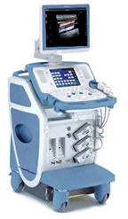
Toshiba Xario Ultrasound
Meet Your Challenging Clinical Demands with Outstanding Performance & Comfort
Excellent diagnostic performance, operator comfort that inspires productivity, and outstanding connectivity features, Xario quite simply, the prime ultrasound system for a wide range of clinical applications.
Below is a generalized description of this systems technology, specifications, features, and options. The below may not reflect the features and options available on units in our inventory.
Xario offers unsurpassed image quality, backed by unique, clinically proven technologies. Its full range of advanced imaging functions lest you visualize minute tissue details and vascular structures with precision, for faster, more accurate diagnosis.
- Pulse Subtraction THI – optimizes spatial and contrast resolution in grayscale imaging. A standard feature with all Xario transducers.
- ApliPure – Real-time compounding delivers images of outstanding clarity and detail, while preserving clinically significant markers.
- Advanced Dynamic Flow – Brings superior spatial resolution to Color Doppler. Depicts even tiny vessels and flow around plaques with improved accuracy and detail.
- Low Ml Pulse Subtraction – Real-time Contrast imaging with outstanding sensitivity in an easy-to- use package for daily practice.
- Trapezoid Imaging – extends the field of view for abetter overview of the region of interest in both grayscale and Color Doppler modes-
- Panoramic View – Reconstructs a single wide-view frame from continuous ultrasound images for improved visualization of widespread regions and anatomical relationships.
- Fast Fusion 3D – Provides clear 3D images of complex structures, such as tumors and their feeding vessels, with simple operation.
- 4D Imaging – Creates surface renderings of structures such as the fetal face with high accuracy and outstanding detail, MPR mode provides three orthogonal scan planes simultaneously.
Ergonomic User Interface
- High-performance TFT Screen with flexible arm and convenient handle.
- Elevating panel adjust quickly to your eyelevel and posture.
- Fully customizable main panel with exchangeable key tops.
- Sensitive touch screen for selecting advanced functions.
QuickScan
- Optimizes 2D gain level instantaneously with acoustic precision.
- Suppresses white noise in echo-weak regions automatically.
- Reduces scan time for a faster, more accurate diagnosis.
- Improves consistency of work flow and overall quality of exams.
Toshiba’s iAssist
- Enables full remote system operation using wireless Bluetooth technology.
- Lessens operator stress by enabling comfortable scanning positions.
- Simplifies complex exams with one—button execution of user-defined protocols.
- Reproduces complex and routine exams using optimum scanning conditions.
System Specifications
Xario with CRT Monitor
- Foot Print – USA (H x W x D) 57.3 – 61.2″ x 21.3″ x 32″
- Foot Print – Metric (H x W x D) 145.5 – 155.5 cm x 54 cm x 81.4 cm
- Weight – 330.8 lbs
Xario with LCD Monitor
- Foot Print – USA (H x W x D) 53.1 — 62.3″ x 21.3″ x 32″
- Foot Print – Metric (H x W x D) 134.8 — 158.3 cm x 54 cm x 81.4 cm
- Weight – 308.7lbs
Monitor
- 17 inch CRT or LCD monitor
- Monitor tilt range: variable positions
- Monitor rotation: variable positions
User Interface
- 450 fps 2D Frame Rate (probe dependant)
- 307 fps Color Frame Rate (probe dependant)
- Pre-Processing YES
- Post-Processing YES
- 1 – 28 cm Display Depth (min/max – probe dependant)
- Selectable Dynamic Range
- Adjustable Transmit Focus
- Customizable Presets
- Gel Warmer
Imaging Modes
- 2D
- M-Mode
Doppler Modes
- High Frame Rate Color Flow
- Color Doppler Velocity
- Color Doppler Energy
- Color Power Doppler
- Directional Tissue Imaging (DTI)
- Pulse Wave (PW)
- Continuous Wave (CW)
Software Technologies
- 3D Imaging
- 4D Imaging
- Compounding
- Speckle Reduction
- Tissue Harmonic Imaging (THI)
- Auto Gain / Optimization
- Tissue Contrast Enhancement (TCE)
- Trapezoid/ Virtual Convex Imaging
- Dual Imaging
- Split Screen
- Duplex
- Triplex
- Clip Function
Connectivity Ports
- USB
- VGA
- SVHS
- S-Video
- Ethernet
- Composite (analog)
- MO Drive
Image File Format
- AVI
- JPG
- TIFF
- DlCOM
On Board/ External Storage
- CD/DVD
- Cine Clips
- Hard Drive 80 GB
- Query & Retrieve
Power Supply
- AC 120 VAC +/- 10%
Peripherals (Options)
- DlCOM
- ECG
- Foot Switch
- Gel Warmer
- Printer
System Applications & Reporting
- Abdomen
- Cardiac Imaging
- Stress Echo
- Obstetrics Imaging
- 3D/4D Imaging
- Contrast Imaging
- General Imaging
- Pediatrics
- Renal
Pain Management Imaging
- Musculoskeletal (MSK)
Women’s Imaging
- Breast Imaging
- Gynecology
- Obstetrics
- 3D/4D
Cardiac Imaging
- Cardiac Screening/ Survey
- Adult Echo
- ECG
- Pediatrics Echo
- Stress Echo
- TEE
Small Parts
- Breast
- Testicle
- Thyroid
Vascular Imaging
- Arterial
- Carotid
- Volume Flow
- Venous
Structured Reporting
- DICOM-Cardiac Structured Reporting
- DICOM- Structured Reporting
Transducers/Probes
- Linear Array 3.1-14 MHz*
- Curved Array 1.9-9.2 MHz
- Phased / Sector Array 2-8.5 MHz*
Cardiac Transducers/ Probes
- Adult Cardiac 2-6 MHz*
- Pediatric Cardiac 2-6.5 MHz*
- Pencil 2 & 5 MHz
- TEE 2.5-7.5 MHz*
3D/ Volume Transducers/ Probes
- Abdominal 2.8-7 MHz
- Endocavity Multifrequency 3.6-8.8 MHz
*Displayed MHz range includes multiple transducers
Transducers:
- PLT 604AT
- PLT 704AT
- PLT 704SBT
- PLT 805AT
- PLT 1202S
- PLT 1204BT
- PVT 375BT
- PVT 382BT
- PVT 674BT
- PVT 712BT
- PVT 661VT
- PVT 770RT
- PST 20CT
- PST 65AT
- PST 50AT
- PST 30BT
- PST 25BT
- PVT 745BTF
- PVT 745BTH
- PVT 67MV
- PVT 575MV
- PVT 382MV
- PVT 375MV
- PET 508MA
- PET 512MC
- PET 511BTM
- PET 510MB
- PC 50M
- PC 20M


