Philips iU22 Ultrasound
A Revolution in Premium Performance Ultrasound
The Philips iU22 xMATRIX ultrasound system is premium performance ultrasound like you’ve never seen before.
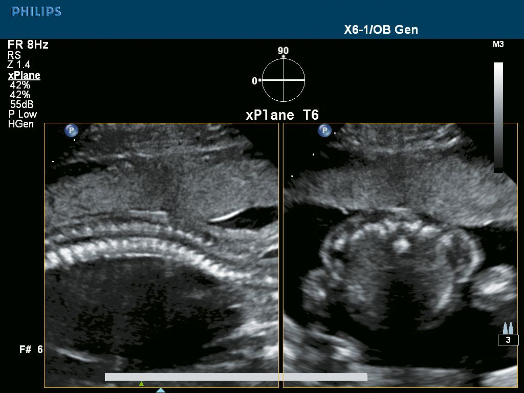
Unique xMATRIX technology on the X6-1 PureWave transducer harnesses the power of over 9,000 active elements, more than 35x greater than today’s conventional transducers, to capture crisp, high-resolution images of even technically challenging patients.
Below is a generalized description of this systems technology, specifications, features, and options. The below may not reflect the features and options available on units in our inventory.
The Philips iU22 xMATRIX ultrasound system is premium performance ultrasound like you’ve never seen before.
Unique xMATRIX technology on the X6-1 PureWave transducer harnesses the power of over 9,000 active elements, more than 35x greater than today’s conventional transducers, to capture crisp, high-resolution images of even technically challenging patients.
System Specifications
- Foot Print- USA (HxWxD) 51-58″ x 21″ x 43.5″
- Foot Print- Metric (HxWxD) 129.5-147.3 cm x 53.3 cm x 110.5 cm
- Weight 220 lbs
User Interface
- Monitor Size 17″LCD Flat Panel
- Programmable Keyboard Buttons
- Moveable Console Variable
- Adjustable Monitor Variable
- Up to 785 fps 2D Frame Rate (probe dependent)
- Pre-Processing
- Post- Processing
- Up to 35cm Display Depth (min/max-probe dependent)
- Selectable Dynamic Range
- Adjustable Transmit Focus
- >100 multiple applications customizable presets
- 2 Transducer Ports
Imaging Modes
- B-Mode (2D)
- M-Mode
Doppler Modes
- High Frame Rate Color Flow
- Color Doppler Velocity
- Color Doppler Energy
- Color Power Doppler
- Directional Color Power Doppler
- directional tissue Imaging (DTI)
- Pulse Wave (PW)
- Continuous Wave (CW)
Software Technologies
- 3D Imaging
- 4D Imaging Option
- Compounding
- Speckle reduction
- Tissue Harmonic Imaging (THI)
- Auto Gain/ Optimization
- Panoramic Imaging
- Dual Imaging
- Split Screen
- Duplex
- Triplex
- Clip Function
Connectivity Ports
- USB
- DVI
- VHS
- ECG
- MO Drive
Image File Format
- AVI
- DICOM
- JPG
Onboard/ External Storage
- CD/DVD
- Cine Clips
- Hard Drive 80 GB
- MO Disk Drive
Power Supply
- AC 100-240 V, 50/60 Hz
Peripherals (Options)
- DICOM
- ECG
- Printer
System Applications & Reporting
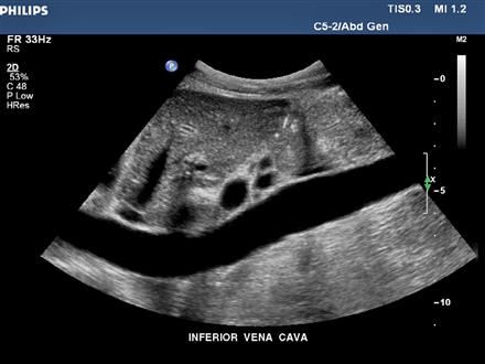
- Abdomen
- General Imaging
- Contrast Imaging
- Intraoperative/ Interventional
- Neonatal
- Pediatrics
- Renal
Pain Management Imaging
- Musculoskeletal (MSK)
Women’s Imaging
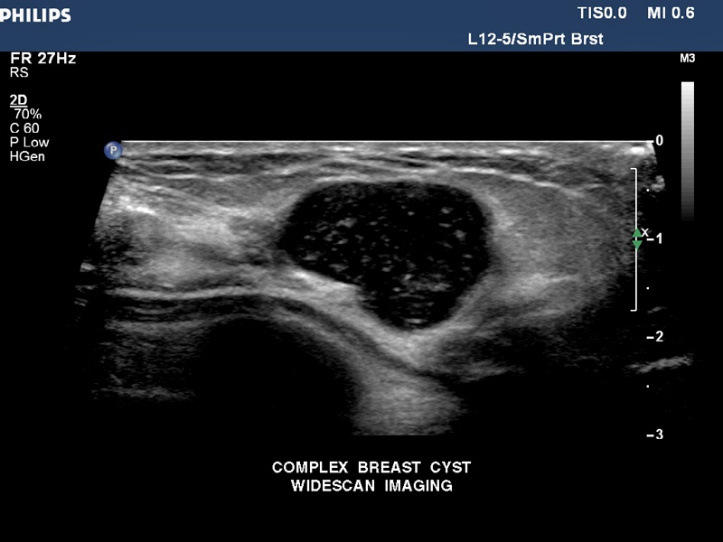
- Breast Imaging
- Gynecology (Option)
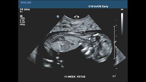
- Obstetrics (Option)
Cardiac Imaging
- Adult Echo
- Cardiac Screening/ Survey
- ECG
- Fetal Echo
- Pediatric Echo
- Stress Echo
- TEE
Small Parts
- Breast
- Testicle
- Thyroid
Urology Imaging
- Prostate
Vascular Imaging
- Transcranial
- Arterial (Option)
- Carotid (Option)
- Venous (Option)
Structured Reporting
- DICOM-Cardiac Structured Reporting
- DICOM- Vascular Structured Reporting
- DICOM- OB/GYN Structured Reporting
User-Programmable Formulas & Tables
Transducers/Probes (*Displayed MHz Range Includes Multiple Transducers)
- Linear Array 3-15 MHz*
- Intraoperative/ Hockey Stick 4-12 MHz
- Curved Array 1-9 MHz*
- Mirco-Convex 5-8 MHz
- Phased/Sector Array 1-12 MHz*
- Veterinary (Equine) 5-8 MHz
Cardiac Transducers/ Probes
- Adult Cardiac 1-12 MHz*
- Pencil 2 MHz & 5 MHz
- TEE 3-8 MHz
- Fetal Cardiac 3-8 MHz
- Pediatric Cardiac 1-12 MHz*
3D/ Volume Transducers/ Probes
- Phased/Sector 1-7 MHz*
- TEE 1-8 MHz*
Transducers: |
|