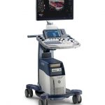
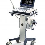
Compact Ultrasound System

The Vivid e delivers everything you need in a compact echo system, like complete cardiovascular functionality. A comprehensive cardiac measurement and analysis package, and full shared service capabilities.

Below is a generalized description of this systems technology, specifications, features and options. The below may not reflect the features and options available on units in our inventory.

Also available used or refurbished. Request a quote for current pricing and availability. See all used and refurbished systems

Transducers/ Probes (*Displayed MHz Range Includes Multiple Transducers)

  • Linear Array 4-12 MHz
  • Intraoperative / Hockey Stick 4-10 MHz
  • Curved Array 2—5.5 MHz
  • Phased / Sector Array 1.5-4 MHz

Cardiac Transducer/ Probe

  • Adult Cardiac 1.5-4 MHz

The Vivid e delivers everything you need in a compact echo system, like complete cardiovascular functionality. A comprehensive cardiac measurement and analysis package, and full shared service capabilities.

  • Comprehensive, diagnostic exams with the conf‌idence building image quality expected from the GE Ultrasound Vivid product line.
  • Easy-to-use, automatic image optimization — in 20, color and Doppler imaging instantly updates thousands of parameters with one keystroke for optimal scanning.
  • Anatomical M-Mode assists with off-axis orientation to make scanning easier.

A Huge Impact on Productivity

  • From how soon the system is ready to scan, to how quickly and efficiently you acquire and store the ultrasound data, the compact Vivid e can make a big difference in your workflow.
  • Pick up the lightweight system. Carry It from room to room.
  • Its rapid power-up and exam pie-sets reduce waiting time to get the images you need.
  • Once you’ve acquired the data, a single press of a button stores images and loops to he onboard 40-GB ard drive so you avoid the delay of storing images to just a memory card.

Measurements and analyses can then be performed whenever you prefer — after the patient has left or at the end of the day. The powerful PC-based platform and large onboard hard drive of the Vivid e make it easy to recall images. So the original, high-quality image is there whenever you need it

Dimensions & Weight

  • Depth: 327 mm (13.35 in)
  • Width: 340 mm (13.88 in)
  • Height 76.5 mm (3.12 in)
  • Weight without battery 4 kg (8.9 lb)
  • Weight with battery 4.5 kg (10 lb)

Console Design

  • Five active probe ports
  • ECG port
  • Integrated HDD
  • Multiple USB ports
  • Integrated DVD-R multi drive
  • On-board DVR recorder (optional)
  • On-board storage for thermal printer
  • Integrated speakers with sub-woofer for premium sound
  • Integrated locking mechanism that provides rolling lock and caster swivel lock
  • Integrated cable management
  • Easily removable air f‌ilters
  • Front and rear handles
  • Side storage trays

User Interface

Operating Keyboard

  • Easy-to-leam user interface with intelligent keyboard
  • Keyboard with appf‌ication-specif‌ic push buttons for primary controls
  • Full-size, alphanumeric keyboard with backlighting
  • Application-specif‌ic secondary controls available through dual rotation and push buttons
  • 6 Slide pot TGC curve
  • Overall gain for 20-mode, Active Mode, on same rotary
  • Digital harvesting of images and loops into image clipboard
  • Patient Browser Screen for registration of demographic data and quick review of image clipboard contents
  • Fully programmable user presets for probe/application default settings
  • Integrated speakers

Display Screen

  • 15″ LCD f‌lat screen
  • Display Pixel: 1024 x 768
  • Screen Tilt Angle: 0 to 160°
  • Wide-angle visibility

Image Management

  • DICOM image format and workflow with instant access data management Images can be transferred in DICOM format to the EchoPac for review.
  • 20 and CFM data at maximum frame rate may be reviewed by scrolling or by running cine loops
  • Image Clipboard for stamp-sized storage and review of stored images and loops
  • Built-in patient archive with images/loops, patient information, measurements and report
  • Internal archive data can be exported to Removable
  • Image Storage through DVD/CD-RW, USB f‌lash card

DICOM Network Connectivity

  • Images transferred in DICOM format to EPPC, alternative workstation or network
  • Wireless LAN connection (option)
  • Ethernet network connection
  • DICOM support
  • Storage to DICOM server
  • Modality Worklist: gives access to a list of patients from a worklist server (usually HIS)
  • Storage commitment
  • Performed procedure step
  • DICOM Print

DICOM Media (Options)

  • Internal Hard Disk: for storing programs, application defaults, ultrasound image and patient archive Over 4OGbyte disk-space for exam archive storage
  • AVI, MPEG and JPEG export
  • DICOM Verification
  • DICOM Print

Peripherals (Options)

  • USB BNV video printer with control from system panel
  • USB color video printer with control from system panel
  • USB inkjet printer
  • USB flash memory card
  • Wireless network interface

Accessories (Options)

  • Replacement battery
  • Replacement hard disk
  • Safety lock
  • Video converter

MPEGvue

  • Using MPEGVue, exams may be stored onto removable media or on remote networked system together with integrated MPEGvue Player for viewing on standard PC

Mobile Cart (Optional)

  • 4 outlet power strip and isolation transformer
  • Probe and gel holder
  • Hand-rest and handles
  • 4 Wheels: 2 swivel, 2 fixed
  • Adjustable peripherals shelves

Electrical Power

  • Battery or mains-line operation
  • AC Adapter Voltage input: 100-240 VAC
  • Frequency: 50/60 Hz
  • Power. Max 130 VA with peripherals

Inputs & Outputs

  • SVGA Video out
  • Audio out
  • USB-2 (to support DVD-RW, video printers, memory stick, etc.)
  • LAN Ethernet
  • USB wireless LAN card
  • DC power input

Analysis Programs

  • Comprehensive Cardiac calculation package
  • Bodymark icons for location and position of probe
  • Vascular measurements package
  • Full M&A Calculation Package with
  • Real Time Auto Doppler Calculations (application specif‌ic)
  • Measurements assignable to protocol capability
  • Parameter annotation follow ASE standard
  • Seamless data storage and report creation
  • Measurements are summarized in worksheets —individual results can be edited or deleted
  • User-assignable parameters
  • Insite-II capability
  • Comprehensive OB package option

System Applications & Reporting

  • Abdomen
  • Contrast Imaging
  • General Imaging
  • Renal

Women Imaging

  • Gynecology
  • Obstetrics

Cardiac Imaging

  • Adult Echo
  • Cardiac Screening/ Survey
  • Stress Echo

Small Parts

  • Breast
  • Testicle
  • Thyroid

Vascular Imaging

  • Arterial
  • Carotid
  • Venous

Request a quote

Fill in your details and a specialist will respond within 24 hours with pricing and availability.

Related models you may also consider

GE Logiq S8

Provide excellent care for your patients with the LOGIQ® S8. We

GE Logiq V1

You use every tool possible to help your patients achieve healthier

GE Vivid T8

The Vivid* T8 combines the established cardiac imaging capabilities of GE