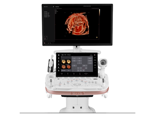
Designed from the ground up with an all-digital architecture, the X150 system provides exceptional performance and instantaneous activation of clinical workflow steps. The X150 system integrates state-of-the-art imaging technologies to address a wider range of patient needs and clinical applications.
Key Technologies Include:
- MultiBeam formation technology increases signal processing speed and delivers exceptional temporal resolution in all imaging modes
- Hanafy lens transducer technology combined with SynAps synthetic aperture technology provides uniformity throughout the field of view up to 28 cm image depth—critical when scanning technically difficult-to-image patients
- Virtual format imaging increases anatomic information by providing a trapezoidal image format at the touch of a button
- All-digital architecture provides signal integrity from beam forming to image display for best-in-class image quality
- MultiHertz multiple frequency imaging technology offers optimized image quality over a wide frequency bandwidth providing exam versatility in a single transducer
System Features:
- Superior 2D-mode imaging- The base system includes superior 2D-mode imaging and pulsed wave Doppler functionality providing high-quality audio and visual capabilities for spectral visualization and quantification of blood flow
- 2D-mode imaging. Color Doppler exams. Cardiac screening. Decide what’s needed — and when.
- Take advantage of the 15-inch, flat panel display mounted on an innovative articulating arm to optimize the position of the system and its folding monitor in virtually any environment
- Configure the transducers and gel holders to fit specific needs
- Customize exam reports to match workflow
- Optimize image quality at the click of a button with TGO tissue grayscale optimization technology
- Quickly store, recall and manage data with the DlMAQ-IP integrated workstation
- Seamlessly integrate the system into networked environments with a full complement of plug-and-play connectivity solutions
Optional Technologies Include:
- Phase inversion Tissue Harmonic Imaging (THI) technology to enhance contrast and spatial resolution and reduce image noise for enhanced visualization across applications
- TGO tissue grayscale optimization technology automatically optimizes the image with the press of a single button, shortening exam times, reducing repetitive motion providing consistent optimized exams across patients, users and applications
- Color and Power Doppler provides superb detection and demonstration of blood flow across all examinations
- 3-Scape real-time 3D imaging provides a real-time reconstruction of 3D images during free hand acquisition
- Cardiac Screening enables phased array imaging capabilities for adult and pediatric cardiac screening
- DlCOM OBGYN Structured Reporting provides standardized report architecture to allow for easy transfer of OB and GYN measurements to offline PCs, workstations and archiving systems
- DlCOM 3.0 Connectivity enables digital data transfer via a DlCOM network for both printing and storage.
- The ACUSON X150 system acts as a DlCOM Storage Class User and DlCOM Print Class User
- DlCOM Modality Worklist enables query and direct download of the patient worklist schedule from the Hospital/Radiology Information System (HIS/RIS) to the ACUSON X150 system, automatically populating the “New Patient‘ screen with patient demographic information
- DlCOM Modality Performed Procedure Step (MPPS)** enables automatic exchange of “Modality Performed Procedure Step“ information with the HlS/RIS
- Additional Array Portadds a third active array port to the ACUSON X150 system and allows the user toconnect up to three linear, curved and phased array transducers simultaneously
* Requires DlCOM 3.0 Connectivity option
**Requires DlCOM 3.0 Connectivity option and DlCOM Modality Worklist option