Philips EPIQ 7 Ultrasound
Breaking Old Rules. Creating New Realities
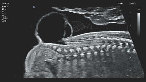
EPIQ 7 is the new direction for premium ultrasound, featuring an uncompromised level of clinical performance to meet the challenges of today’s most demanding practices and technically difficult-to-image patients through every gestational age and for gynecology applications.
The Philips proprietary nSIGHT Imaging architecture introduces a totally new approach to forming ultrasound images without compromise.
Below is a generalized description of this systems technology, specifications, features, and options. The below may not reflect the features and options available on units in our inventory.
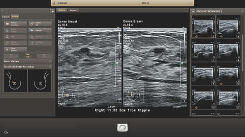
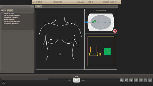
Philips AI Breast is an integrated solution for whole breast ultrasound. AI Breast offers screening, diagnostic, and workflow benefits utilizing Philips unique Anatomical Intelligence. Designed with both the user and patient in mind, AI Breast allows the ultrasound scan room to be utilized for a full range of examinations without additional obtrusive hardware.
Philips exclusive PureWave crystal technology is clinically proven to improve penetration in difficult-to-image patients. The pure, uniform PureWave crystals are up to 85% more efficient than conventional materials, resulting in exceptional performance. This technology allows for improved penetration and excellent detailed resolution.
Philips proprietary nSIGHT Imaging architecture is a totally different approach to forming ultrasound images. Unlike conventional systems that form the image line by line, nSIGHT creates images with optimal resolution down to the pixel level. nSIGHT Imaging incorporates the use of a precision beamformer along with powerful massive parallel processing. This extraordinary architecture captures an enormous amount of acoustic data and then reconstructs in real time optimally focused beams, creating precise resolution for every pixel in the image.
EPIQ’s architecture supports the Philips exclusive Anatomical Intelligence Ultrasound (AIUS), designed to elevate the ultrasound system from a passive to an actively adaptive device. With advanced organ modeling (with xMATRIX technology), and proven quantification, exams are easy to perform, more reproducible, and deliver new levels of clinical information. AIUS ranges from automating repetitive steps to full, computer-driven analysis with minimal user interaction – all using anatomic intelligence and all providing the results you need.
Make confident decisions even in challenging diagnostic cases with new fully integrated fusion capabilities that feature streamlined workflow to allow clinicians to achieve fast and effective fusion of CT/MR/PET with live ultrasound. By combining imaging modalities directly on the ultrasound system, you now have access to an even more powerful diagnostic tool with advanced visualization allowing for fast clinical decisions.
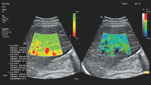
Simplify liver assessment with non-invasive tools. Obtaining liver stiffness measurements with Philips shear wave elastography is surprisingly fast and easy even on difficult-to-image patients. It is non-invasive, making it a quick, simple step for sonographers and virtually painless for patients.
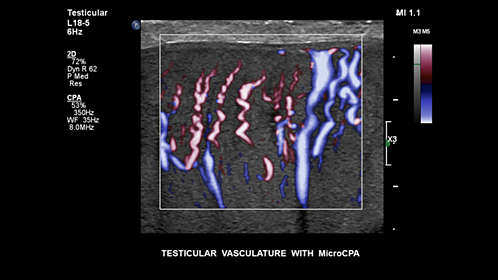
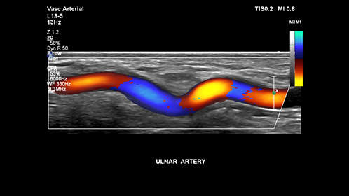
Obtaining flow information in small low-flow vascular structures has traditionally been a challenge. With EPIQ’s new MicroCPA feature, visualization of low velocity micro circulation is quick and simple, giving you more diagnostic confidence when evaluating organ perfusion or small vascular beds.
Real Time iSCAN (AutoSCAN) automatically optimizes gain and TGC to continuously provide a high-quality image.
EPIQ 7 has completely reinvented the premium ultrasound user experience. Ease of use, workflow, ergonomics, and mobility. We’ve revolutionized how you interact with an ultrasound system from every standpoint, and kept it beautifully intuitive and very quiet.
Navigate quickly to system functions with the tablet-like touch interface, with 40% less reach and 15% fewer steps to complete an exam.
EPIQ’s extended-range control panel and monitor can be articulated for proper ergonomic alignment whether sitting or standing. The large 21″ wide screen monitor facilitates easy viewing in virtually any environment. EPIQ 7 has four transducer connectors with ambient lighting for ease in transducer selection during an exam.
The EPIQ ultrasound machine is the lightest in its class; it’s easily transported on both carpet and tile. Place it in sleep mode, move it and boot up in seconds. The monitor folds down to reduce overall system height for transport, and the integrated cable hooks and catch tray are ideal for mobile studies.
Philips TrueVue advanced 3D ultrasound display delivers amazing lifelike 3D images. TrueVue, with its internal light source gives clinicians the ability to manipulate light and shadow anywhere in the 3D volume.
View DICOM images such as CT, NM, MRI, mammography, and ultrasound on your EPIQ system. Easily compare past and current studies without the use of an external reading station, and even review these Multimodality images while live imaging. Capture side-by-side comparison images as part of the exam documentation.
EPIQ 7 is almost silent when running A noise test determined that EPIQ 7 runs at 37-41 dB, which is equivalent to the sound of a library. This is extremely welcome in small scanning/examination rooms.
At the touch of a button, MaxVue high-definition display brings extraordinary visualization of anatomy with 1,179,648 additional image pixels compared to a standard4:3 display format mode. MaxVue enhances ultrasound viewing and provides 38% more viewing area to optimize the display of dual, side/side, biplane, and scrolling imaging modes.
System Specifications
- Foot Print- Metric (HxWxD) 146-171.5 cm x 60.6 cm x 109.2 cm
- Weight- 230 lbs
Control Panel
- Moniter Size- 54.6 cm
- Touch Screen Size- 12″
- Height Adjustment- 25.4 cm
- Degrees of Movement- 180 degrees
User Interface
- Monitor Size 17″LCD Flat Panel
- Monitor Resolution- 1920×1080
- Programmable Keyboard Buttons
- Moveable Console Variable
- Adjustable Monitor Variable
- Up to 785 fps 2D Frame Rate (probe dependent)
- Pre-Processing
- Post- Processing
- Up to 35cm Display Depth (min/max-probe dependent)
- Selectable Dynamic Range
- Adjustable Transmit Focus
- >100 multiple applications customizable presets
- 2 Transducer Ports
Imaging Modes
- B-Mode (2D)
- M-Mode
- 3D/4D
Doppler Modes
- High Frame Rate Color Flow
- Color Doppler Velocity
- Color Doppler Energy
- Color Power Doppler
- Directional Color Power Doppler
- directional tissue Imaging (DTI)
- Pulse Wave (PW)
- Continuous Wave (CW)
Software Technologies
- 3D Imaging
- 4D Imaging Option
- Compounding
- Speckle reduction
- Tissue Harmonic Imaging (THI)
- Auto Gain/ Optimization
- Panoramic Imaging
- Dual Imaging
- Split Screen
- Duplex
- Triplex
- Clip Function
Connectivity Ports
- USB
- DVI
- VHS
- MO Drive
Image File Format
- AVI
- DICOM
- JPG
Onboard/ External Storage
- CD/DVD
- Cine Clips
- Hard Drive 80 GB
- MO Disk Drive
Power Supply
- AC 100-240 V, 50/60 Hz
Peripherals (Options)
- DICOM
- ECG
- Printer
System Applications & Reporting
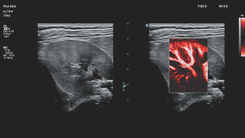
- Abdomen
- General Imaging
- Contrast Imaging
- Intraoperative/ Interventional
- Neonatal
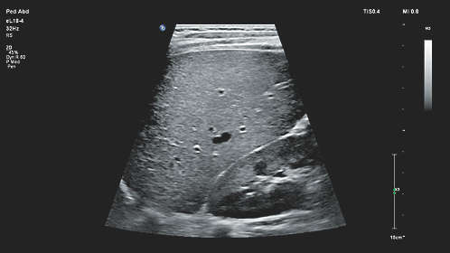
- Pediatrics
- Renal
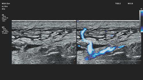
Pain Management Imaging
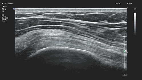
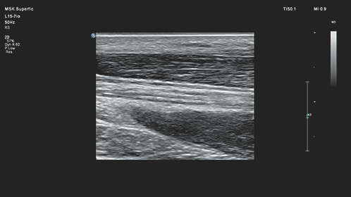
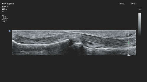
- Musculoskeletal (MSK)
Women’s Imaging
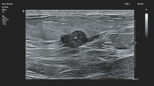
- Breast Imaging
- Gynecology
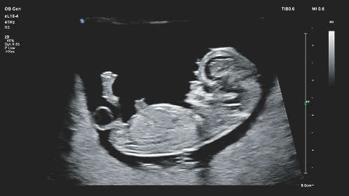
- Obstetrics
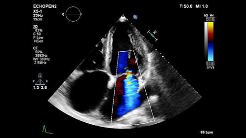
Cardiac Imaging
- Adult Echo
- Cardiac Screening/ Survey
- ECG
- Fetal Echo
- Pediatric Echo
- Stress Echo
- TEE
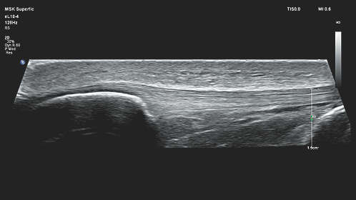
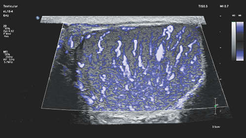
Small Parts
- Breast
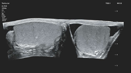
- Testicle
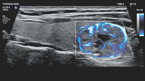
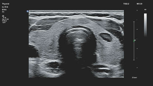
- Thyroid
Urology Imaging
- Prostate
Vascular Imaging
- Transcranial
- Arterial
- Carotid
- Venous
Structured Reporting
- DICOM-Cardiac Structured Reporting
- DICOM- Vascular Structured Reporting
- DICOM- OB/GYN Structured Reporting
User-Programmable Formulas & Tables
Curved Transducers/ Probes
- C5-1 (1-5 MHz)
- C9-2 (2-9 MHz)
- C8-5 (5-8 MHz) – Mirco
- V6-2 (2-6 MHz) – 3D
- VL13-5 (5-13 MHz) – 3D
Linear Transducers/ Probes
- L12-3 (3-12 MHz)
- L12-5 50mm (5-12 MHz)
- L18-5 (5-18 MHz)
- L15-7io (7-15 MHz) – Hockey
Phased Transducers/ Probes
- S5-1 (1-5 MHz)
- S8-3 (3-8 MHz)
- S12-4 (4-12 MHz)
- X5-1 (1-5 MHz)
- X6-1 (1-6 MHz)
Endocavity Transducers/ Probes
- 3D9-3V (3-9 MHz)
- C10-3V (3-10 MHz)
- C10-4ec (4-10 MHz)
Pencil (CW) Transducers/ Probes
- D2tcd PW (2 MHz)
- D5cwcCW (5 MHz)
- D2cwc CW (2 MHz)
Transesophageal Transducer/ Probe
- X7-2t (2-7 MHz)