Philips HD11 Ultrasound
High-Definition Imaging in a Complete Package
Ready to expand with your clinical needs, the HD11 XE is a complete digital imaging system that delivers high-definition imaging and ease-of-use in a compact, ergonomic, and reliable package.
Below is a generalized description of this systems technology, specifications, features, and options. The below may not reflect the features and options available on units in our inventory.
The HD11 XE is ready to expand with your clinical needs. It is a complete, digital imaging system that delivers high-definition imaging and ease-of-use in a compact, ergonomic, and reliable package.
This system has the versatility to expand into a complete, digital cardiovascular imaging system for further clinical utility and value. You can also add powerful options like QLAB advanced quantification, stress echo, contrast imaging, and transesophageal echo to further enhance your cardiovascular imaging capabilities.
Get outstanding image quality and ease-of-use for ultrasound guided regional anesthesia and emergency medicine with special configurations that are surprisingly affordable.
The HD11 XE brings 4D imaging into mainstream by delivering it on a platform with an unmatched combination of versatility and value. Move seamlessly through 2D and Doppler modes right into breathtaking 4D studies. The system’s powerful architecture supports continuous, precise, quantitative volume acquisition and display, with easy, simultaneous visualization and measurements in three planes. Numerous innovations are on-and-off-cart to give you stunning image quality and the potential to improve your exam efficiencies by changing the way you acquire and visualize ultrasound data.
- SonoCT compounding
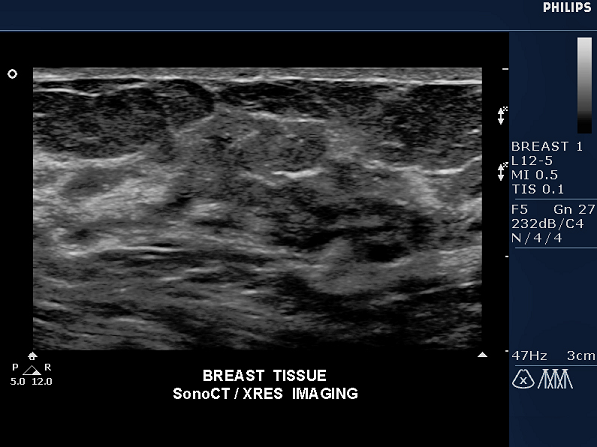
- XRES processing to eliminates speckle noise artifact
- 2D with Pulse Inversion Harmonic Imaging
- 3D imaging with multiplanar views, for qualitative freehand 3D images
- Adaptive Color Doppler
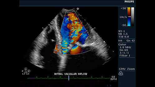
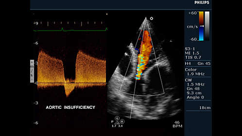
- Pulsed wave and continuous wave Doppler with adaptive Doppler technology
The contrast option equips the HD11 XE to detect harmonic agent signatures using the S3-1 and C5-2 transducers. The option provides a uniform power field, allowing more even excitement of contrast agents throughout the sector. Optimized LVO system settings on the HD11 XE decreases contrast agent destruction and increase ease-of-use by minimizing the need for system adjustments. All of which add up to more complete visualization of contrast throughout imaging.
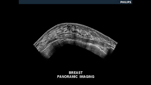
The Panoramic Imaging option provides an extended field-of-view display. This feature creates a series of real-time images while the user moves the transducer laterally across the anatomy. When the imaging is complete, the system renders a panoramic mosaic display. The resulting panoramic image provides a larger reference image for documentation of spatial relationships of structure.
Image and data management capabilities allow flexible recording, archiving, editing, and even exam reports with embedded images.
Multi session CD and optional peripheral device allow you to meet your documentation and archiving needs. Prepare professional patient reports with embedded images.
System Specifications
- Foot Print- USA (HxWxD) 51-58″ x 21″ x 43.5″
- Foot Print- Metric (HxWxD) 129.5-147.3 cm x 53.3 cm x 110.5 cm
- Weight 220 lbs
User Interface
- Monitor Size 17″LCD Flat Panel
- Programmable Keyboard Buttons
- Moveable Console Variable
- Adjustable Monitor Variable
- Up to 785 fps 2D Frame Rate (probe dependent)
- Pre-Processing
- Post- Processing
- Up to 35cm Display Depth (min/max-probe dependent)
- Selectable Dynamic Range
- Adjustable Transmit Focus
- >100 multiple applications customizable presets
- 2 Transducer Ports
Imaging Modes
- B-Mode (2D)
- M-Mode
Doppler Modes
- High Frame Rate Color Flow
- Color Doppler Velocity
- Color Doppler Energy
- Color Power Doppler
- Directional Color Power Doppler
- directional tissue Imaging (DTI)
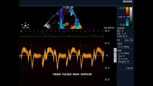
- Pulse Wave (PW)
- Continuous Wave (CW)
Software Technologies
- 3D Imaging
- 4D Imaging Option
- Compounding
- Speckle reduction
- Tissue Harmonic Imaging (THI)
- Auto Gain/ Optimization
- Panoramic Imaging
- Dual Imaging
- Split Screen
- Duplex
- Triplex
- Clip Function
Connectivity Ports
- USB
- DVI
- VHS
- ECG
- MO Drive
Image File Format
- AVI
- DICOM
- JPG
Onboard/ External Storage
- CD/DVD
- Cine Clips
- Hard Drive 80 GB
- MO Disk Drive
Power Supply
- AC 100-240 V, 50/60 Hz
Peripherals (Options)
- DICOM
- ECG
- Printer
System Applications & Reporting
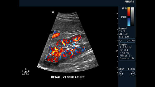
- Abdomen
- General Imaging
- Contrast Imaging
- Intraoperative/ Interventional
- Neonatal
- Pediatrics
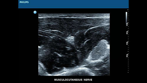
Pain Management Imaging
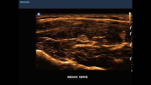
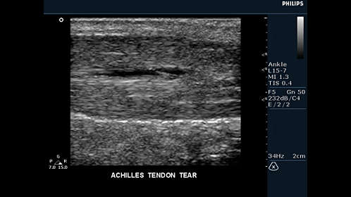
- Musculoskeletal (MSK)
Women’s Imaging
- Breast Imaging
- Gynecology
- Obstetrics
- Reproductive Medicine (IVF)
Cardiac Imaging
- Adult Echo
- Cardiac Screening/ Survey
- ECG
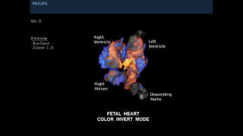
- Fetal Echo
- Neonatal Echo
- Pediatric Echo
- Stress Echo
- TEE
Small Parts
- Breast
- Testicle
- Thyroid
Urology Imaging
- Prostate
- Pelvis
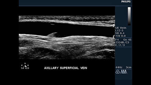
Vascular Imaging
- Transcranial
- Arterial
- Carotid
- Venous
Structured Reporting
- DICOM-Cardiac Structured Reporting
User-Programmable Formulas & Tables
Transducers/Probes (*Displayed MHz Range Includes Multiple Transducers)
- Linear Array 3-12 MHz*
- Intraoperative/ Hockey Stick 7-15 MHz
- Curved Array 2-9 MHz*
- Mirco-Convex 5-8 MHz
- Phased/Sector Array 1-12 MHz*
- Endocavity 5-9 MHz
- Endovaginal 4-9 MHz
- Bi-Plane Endorectal 5-10 MHz
- Veterinary (Equine) 5-8 MHz
Cardiac Transducers/ Probes
- Adult Cardiac 1-12 MHz*
- Pencil 2 MHz & 5 MHz
- TEE 2-7 MHz
- Fetal Cardiac 3-12 MHz
3D/ Volume Transducers/ Probes
- Abdominal 2-8 MHz*
- Endocavity 3-9 MHz*
Transducers:
- L15-7io
- L12-5
- L12-3
- L9-3
- L8-4
- C9-5ec
- C9-4
- C8-5
- C8-4v
- C6-3
- C5-2
- S12-4
- S8-3
- S7-3t
- S7-2t
- S4-2
- S3-1
- V8-4
- V6-2
- 3D9-3v
- 3D8-4
- 3D6-2
- BP10-5ec
- D2cwc
- D2tcd
- D5cwc