Rapid Patient Care

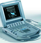
The SonoSite Titan ultrasound is a high-resolution modular portable solution that provides health care professionals with state-of-the-art high-resolution imaging wherever needed for responsive, rapid patient care.

Below is a generalized description of this systems technology, specifications, features, and options. The below may not reflect the features and options available on units in our inventory

Also available used or refurbished. Request a quote for current pricing and availability. See all used and refurbished systems

All transducers are lightweight with rapid, pinless connectors and feature a user selectable menu to adjust frequency settings to suit the patients size. Settings are Penetration, General and Resolution imaging.

Transducers/ Probes * (Displayed MHz Range Includes Multiple Transducers)

  • Linear Array 5-13 MHz*
  • lntraoperative / Hockey Stick 5-10 MHz*
  • Curved Array 2-5 MHz
  • Micro-Convex 5-8 MHz
  • Endovaginal 5-8 MHz
  • Endorectal 5-8 MHz

Cardiac Transducers/ Probes:

  • Adult Cardiac 2-4 MHz

Transducers:

  • L25
  • L38
  • HST
  • C11
  • C15
  • C60
  • C8
  • ICT

The SonoSite® TITAN is designed for those who require the features and functionality of a traditional ultrasound system, but the mobility and durability that only SonoSite can deliver. The innovative TITAN system offers the amazing image quality and technical capabilities of cart-based machines with the ease and flexibility of a hand-carried system.

Amazing Imaging Capabilities.

Advanced technologies, including pulsed wave Doppler, directional color power Doppler, Tissue Harmonic Imaging and M-mode, make TITAN suitable for a wide range of clinical applications. Its all-digital LCD screen displays high-resolution images when and where you need them, improving diagnostic confidence and providing better control for image-guided procedures, resulting in improved patient care.

Imaging Software

  • SonoCalc™ IMT automated edge detection software for assessment of intima media thickness
  • SiteLink image management software (DICOM and Non-DICOM).

System Specifications

  • System weight: 7.7 lbs
  • System dimensions: 11.9″ L x10.9″Wx 3″ H
  • Display: Integrated fully digital 8.4″ (diagonal) TFT
    color Liquid Crystal Display

Imaging Modes and Processing

  • 2D
  • Split screen
  • Zoom
  • Color power Doppler
  • M-Mode
  • 3 sweep speeds, 1/2 1/2, 1/3 2/3 or full screen
    options
  • Pulsed wave (PW) Doppler
  • Continuous wave (CW) Doppler
  • Velocity based color f‌low Doppler
  • Duplex imaging
  • 3 sweep speeds, 1/2 1/2, 1/3 2/3 or full screen
    opt‌ions
  • Tissue Harmonic Imaging (THI)

Mounting Options

  • Mobile Docking System dimensions: 31.9″ L x 22.6″
    W x adjustable height 33.2″ to 45.1″
  • Storage available for fully integrated printer, DVD
    and/or VCR storage area
  • Optional connector for 3 transducers — changes by
    simple electronic switching
  • Optional 11.8″ (diagonal) monitor

User Interface

Programmable Controls

  • Remappable Controls – Softkeys to drive advanced features. Delta key: user selectable functions can be assigned to the delta key for increased ease of use

Measurement Tools

  • 2D
  • Distance calipers — two sets
  • Ellipse for area and circumference measurements
    M-mode
  • Distance and time caliper set
  • Heart rate
  • Doppler
  • Velocity caliper— two sets provide peak velocity RI
    & S/D ratio.
  • Volume Flow

Display Annotation

  • Text and pictograms
  • User selectable application specific text via function keys.
  • Image Storage and Cine
  • Storage: High capacity using f‌lash card. 64MB
    card stores 128 images (approx).
  • Cine: Review of up to 220 frame—by-frame
    images.
  • Connect directly to a PC with SiteLink Image
    Manager software using a USB cable from the
    system. Allows transfers of high resolution
    bitmap images.
  • Software allows batch compression to JPG on
    PC.
  • Image transfer can be active while live scanning
    on TITAN

Software (Post-Processing)

  • SonoCalc IMT automated edge detection software for assessment of intima media thickness

Peripheral Connections

  • Composite video output (NTSC/PAL) to video cassette recorder, video printer or external monitor.
  • Connect to a PC via USB to download high resolution bitmap images.
  • S-video output
  • Ethernet output
  • VGA output

Connectivity

  • DICOM print and storage class software available
  • DICOM worklist

Power

  • Operates via battery or AC power— from 1.5 up to 4 hours on fully charged battery,
  • New battery life depends on the active functions used and keystrokes.
  • Battery: Rechargeable lithium ion
  • AC: Via universal power adapter, 100-240 VAC, 50- 60 Hz

System Applications & Reporting

  • Abdomen
  • Emergency Medicine
  • General Imaging
  • Renal

Pain Management Imaging

  • Musculoskeletal (MSK)
  • Anesthesia
  • Orthopedic

Women’s Imaging

  • Breast Imaging
  • Gynecology
  • Obstetrics

Cardiac Imaging

  • Cardiac Screening/ Survey

Small Parts

  • Breast
  • Testicle
  • Thyroid

Urology Imaging

  • Pelvis
  • Prostate

Vascular Imaging

  • Arterial
  • Carotid
  • IMT
  • Volume Flow
  • Venous

Structured Reporting

  • DICOM-Vascular Structured Reporting
  • DICOM-OB/GYN Structured Reporting

Request a quote

Fill in your details and a specialist will respond within 24 hours with pricing and availability.

Related models you may also consider

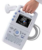
SonoSite 180 Plus

The SonoSite 180 Plus ultrasound provides a sensitive and accurate quantitative
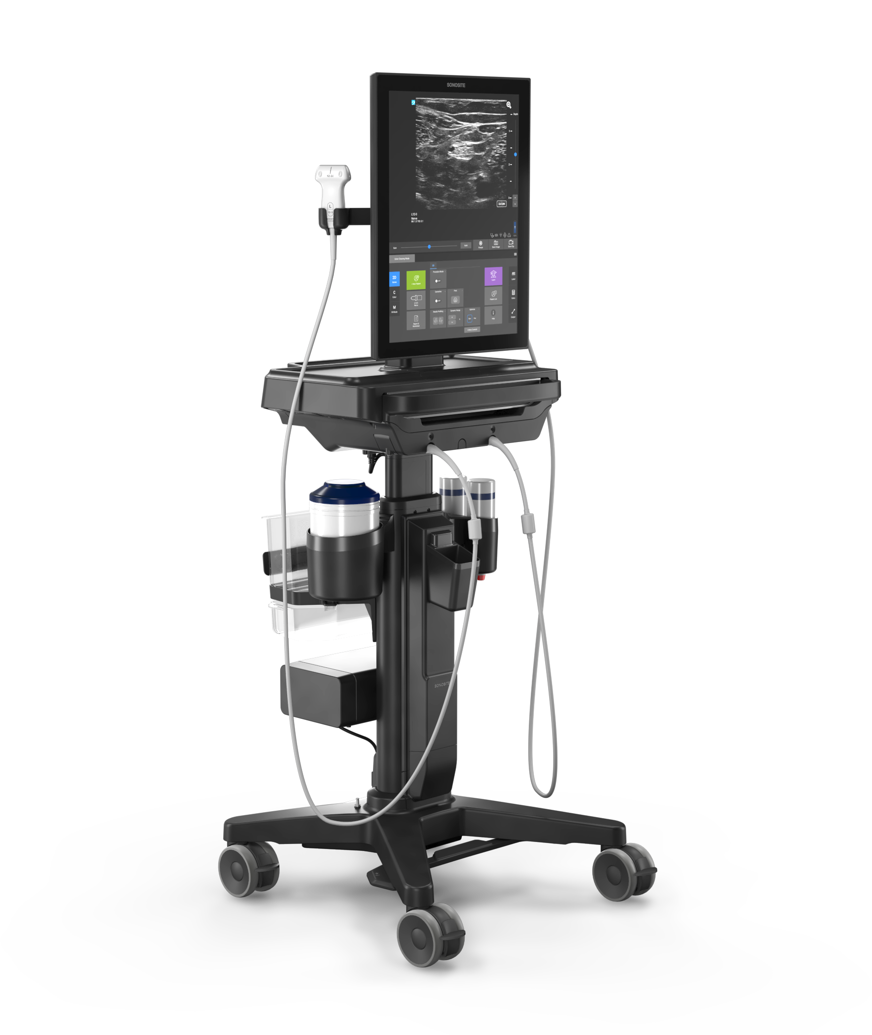
Sonosite ST point-of-care ultrasound cart system featuring 21-inch touchscreen display and sleek ergonomic footprint

FUJIFILM Sonosite ST

The Sonosite ST delivers advanced ultrasound imaging, streamlined workflows, and precise

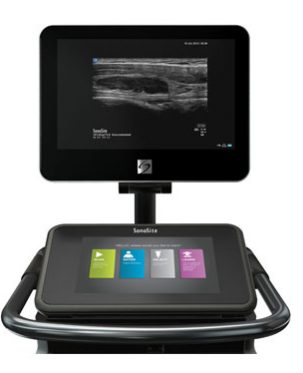
SonoSite X-Porte

X-Porte represents an entirely new approach to clinical ultrasound. Its imaging,