Philips EPIQ 5 Ultrasound
A New Era in Premium Ultrasound
EPIQ 5 is the new direction for premium ultrasound, featuring an uncompromised level of clinical performance to meet the challenges of today’s most demanding practices and technically difficult-to-image patients through every gestational age and for gynecology applications.
Below is a generalized description of this systems technology, specifications, features, and options. The below may not reflect the features and options available on units in our inventory.
EPIQ 5 has completely reinvented the premium ultrasound user experience. Ease of use, workflow, ergonomics, and mobility. We’ve revolutionized how you interact with an ultrasound system from every standpoint, and kept it beautifully intuitive and very quiet.
System Specifications
- Foot Print- Metric (HxWxD) 146-171.5 cm x 60.6 cm x 109.2 cm
- Weight- 230 lbs
Control Panel
- Moniter Size- 54.6 cm
- Touch Screen Size- 12″
- Height Adjustment- 25.4 cm
- Degrees of Movement- 180 degrees
User Interface
- Monitor Size 17″LCD Flat Panel
- Monitor Resolution- 1920×1080
- Programmable Keyboard Buttons
- Moveable Console Variable
- Adjustable Monitor Variable
- Up to 785 fps 2D Frame Rate (probe dependent)
- Pre-Processing
- Post- Processing
- Up to 35cm Display Depth (min/max-probe dependent)
- Selectable Dynamic Range
- Adjustable Transmit Focus
- >100 multiple applications customizable presets
- 2 Transducer Ports
Imaging Modes
- B-Mode (2D)
- M-Mode
- 3D/4D
Doppler Modes
- High Frame Rate Color Flow
- Color Doppler Velocity
- Color Doppler Energy
- Color Power Doppler
- Directional Color Power Doppler
- directional tissue Imaging (DTI)
- Pulse Wave (PW)
- Continuous Wave (CW)
Software Technologies
- 3D Imaging
- 4D Imaging Option
- Compounding
- Speckle reduction
- Tissue Harmonic Imaging (THI)
- Auto Gain/ Optimization
- Panoramic Imaging
- Dual Imaging
- Split Screen
- Duplex
- Triplex
- Clip Function
Connectivity Ports
- USB
- DVI
- VHS
- MO Drive
Image File Format
- AVI
- DICOM
- JPG
Onboard/ External Storage
- CD/DVD
- Cine Clips
- Hard Drive 80 GB
- MO Disk Drive
Power Supply
- AC 100-240 V, 50/60 Hz
Peripherals (Options)
- DICOM
- ECG
- Printer
System Applications & Reporting
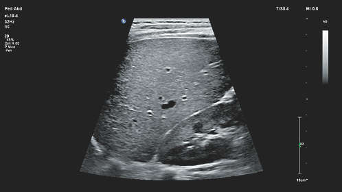
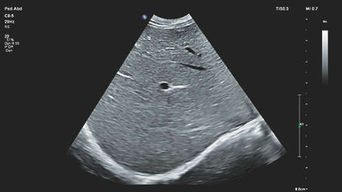
- Abdomen
- General Imaging
- Contrast Imaging
- Pediatrics
- Renal
Pain Management Imaging
- Musculoskeletal (MSK)
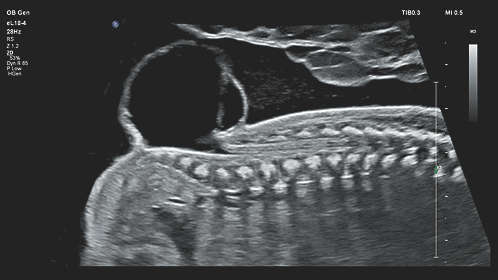
Women’s Imaging
- Breast Imaging
- Gynecology
- Obstetrics
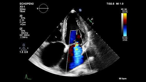
Cardiac Imaging
- Adult Echo
- Cardiac Screening/ Survey
- Pediatric Echo
- TEE
Small Parts
- Breast
- Testicle
- Thyroid
Urology Imaging
- Prostate
Vascular Imaging
- Transcranial
- Arterial
- Carotid
- Venous
Structured Reporting
- DICOM-Cardiac Structured Reporting
- DICOM- Vascular Structured Reporting
- DICOM- OB/GYN Structured Reporting
User-Programmable Formulas & Tables
Curved Transducers/ Probes
- C5-1 (1-5 MHz)
- C9-2 (2-9 MHz)
- C8-5 (5-8 MHz) – Mirco
- V6-2 (2-6 MHz) – 3D
- VL13-5 (5-13 MHz) – 3D
Linear Transducers/ Probes
- L12-3 (3-12 MHz)
- L12-5 50mm (5-12 MHz)
- L18-5 (5-18 MHz)
- L15-7io (7-15 MHz) – Hockey
Phased Transducers/ Probes
- S5-1 (1-5 MHz)
- S8-3 (3-8 MHz)
- S12-4 (4-12 MHz)
Endocavity Transducers/ Probes
- 3D9-3V (3-9 MHz)
- C10-3V (3-10 MHz)
- C10-4ec (4-10 MHz)
Pencil (CW) Transducers/ Probes
- D2tcd PW (2 MHz)
- D5cwcCW (5 MHz)
- D2cwc CW (2 MHz)
Transesophageal Transducer/ Probe
- X7-2t (2-7 MHz)