Mindray M8 Elite Ultrasound
Voice Activated
The Mindray M8 Elite portable ultrasound machine is based on Mindray’s new generation ultrasound platform, mQuadro. Its advanced signal transmission and reception processors provide highly sensitive and accurate echo detection.
The M8 Elite has raised industry standards to an all new high!
Below is a generalized description of this systems technology, specifications, features, and options. The below may not reflect the features and options available on units in our inventory.
The M8 Elite is powered by mQuadro, a new and innovative imaging architecture that incorporates powerful, high speed digital signal processing and intelligent software algorithms that take ultrasound imaging to new levels.
- More rapid image acquisition with fewer keystrokes
- Better image uniformity from near to far field
- Designed for deeper penetration
- Increased exam efficiency and improved patient throughput
- Better signal-to-noise ratio (SNR)
- Use of higher frequencies to image deeper structures
- Easier and faster acquisition of images across all body types
- Better image clarity across all body types
Imaging Modes:
- 2D
- 3D/4D
Applications:
- Abdominal
- Anesthesia
- Breast
- Cardiac
- Emergency Medicine
- General Imaging
- Intraoperative
- MSK
- OB/GYN
- Pediatrics
- Neonatal
- TCD
- Small Parts
- Superficial
- Vascular
- Venous
- TEE
Features:
- M-Mode
- Color M-Mode
- PW Doppler
- CW Doppler
- Color Doppler
- Power Doppler
- DICOM
- Auto Optimization
- Speckle Reduction
- Compound Imaging
- Tissue Harmonics
- HPRF,
- Panoramic
- Needle Visualization
- IMT
- Stress Echo
- Anatomical M-Mode
- Tissue Doppler
Video & Output Options:
- S-Video
- Ethernet
- Audio
- USB
- Composite Video
- DVI
Monitor Resolution:
- 1024×768
Dimensions:
- 14 lbs, 14″ x 14″ x 3″
Export Options:
- DICOM
- JPG
- BMP
- RAW
- USB
USB Ports:
- 2
DICOM Options:
- Store
- Worklist
- MPPS
- Structured Reports
- Query/Retrieve
PC Export Formats:
- AVI
- JPG
- BMP
Wireless:
- Yes
Power (USA):
- 100-240V, 50/60 Hz
HDD Size:
- 160GB
Max Cine Memory:
- 8830 Frames
Maximum Depth (cm):
- 30
Max Frame Rate (FPS):
- 643
Accessories:
- Printer
- Footswitch
- DVD-Recorder
- Cart
- Second Battery
- Wireless Adapter
- VCR
- CD/DVD R/W
Specialty applications:
- Focused Assessment with Sonography in Trauma (FAST)
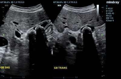
- Gallbladder
- Abdominal Aortic Aneurysm (AAA)
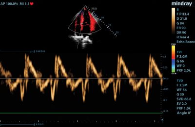
- Cardiology
- Focused cardiac
- Foreign bodies
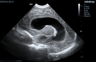
- OB/GYN
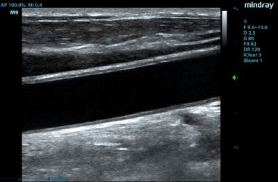
- DVT (Deep Venous Thrombosis)
- Renal evaluations
- Vascular access
- Renal evaluations
- Vascular access
- Line placement
Anesthesia:
- Ultrasound-assisted procedural guidance applications
- Peripheral Nerve Blocks
- lnterscalene
- Supraclavicular
- lnfraclavicular
- Musculocutaneous
- llioinguinal/lliohyposgastric
- Femoral
- Sciatic
- Popliteal
- Saphenous
- Epidural/Spinal
- Vascular Access
Cardiac
- Stress Echocardiograohy:
- Fast, intuitive, user friendly work flow
- 13 factory default stress protocols covering: Exercise, Dobutamine, and Ergometer
- Unlimited user defined protocols with up to 12 stages per study
- Pause function
- Easy global clip edit function
- Wall motion scoring protocols ASE 16, ASE 17
Transesophgeal Echo:
- Motor driven l Forward/reverse and lateral flexion l 180 degree scan plane I 180 degree scan plane indicator
Vascular Access
- Easily connects to your work station, giving you the ability to access essential patient reporting directly on the ultrasound system
- Access to direct applications as diverse as inputting data and editing the patient report from the workstation through the M7 while never leaving the patient’s side
- Provides immediate connectivity, bringing the user in touch with patient data streams such as electronic patient reports, at a single location, without compromising the workflow
Vascular Imaging Applications:
- Carotid exam
- Upper and lower extremity arterial exam
- Venous duplex
- Vascular Access
- Line placement
- TDC
- ABD vascular
- Comprehensive reporting
Curved Transducers/ Probes:
- C5-2s
- 6C2s (Micro)
- 4CD4s (4D)
Linear Transducers/ Probes:
- L14-6s
- L14-6ns
- 7L4s
- L7-3s
- L12-4s
- 7LT4s
Phased Transducers/ Probes:
- P4-2s
- P7-3s
- P12-4s
Endocavity Transducers/ Probes:
- V10-4bs
- V10-4s
CW (Pencil) Transducers/ Probes:
- CW2s
- CW5s
Transesophegeal Transducer/ Probe:
- P7-3ts