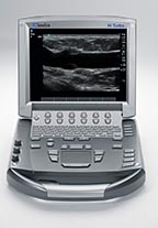
SonoSite M-Turbo Ultrasound
See How Versatile Ultrasound Can Be
The SonoSite M-Turbo portable ultrasound is SonoSite’s nost versatile system for abdominal, nerve, vascular, cardiac, venous access, pelvic, and superficial imaging. The M-Turbo ultrasound tool comes with SonoGT and SonoHD imaging technology, enhanced color flow, optional SonoRemote control.
The M-Turbo has 16x the processing power of our previous generation and still weighs under 7 pounds. As always, it comes with an unparalleled 5-year warranty.
Below is a generalized description of this systems technology, specifications, features, and options. The below may not reflect the features and options available on units in our inventory.
SonoGT™ Global Targeted technology, an advance that capitalizes on the power of the M-Turbo platform to drive a new level of color flow imaging, wireless connectivity and workflow integration for anesthesia, emergency medicine, critical care, and other acute point-of-care markets.
ColorHD Technology – A proprietary, color Doppler algorithm available on all transducers, ColorHD™ technology works in parallel with multiple M-Turbo algorithms, including SonoHD™ imaging technology, to provide increased diagnostic information and better visualization of color flow, particularly in low flow states.
An option using 802.11b/g technology, SonoRoam™ technology enables wireless image transfer from the M-Turbo system to a PACS system via DICOM® and to a personal computer via SonoSite’s SiteLink™ so that clinicians can quickly retrieve the information from any location.
- DICOM Storage Commit – facilitates seamless clinical integration of ordering, scheduling, image acquisition, storage, viewing, and billing of patient procedures.
- Patient Management – Patient demographics can now be entered before, during, or after the exam
- New USB Bar Code Reader – allows clinicians to quickly and accurately enter patient information and update exam information prior to its entry into the patient record
System Specifications
- Weight: 6.7 lbs (3-04 kg) (without battery and transducer)
- Dimensions: 11.9″ L x 10.8″ W x 3.1″ H (30.2 cm L x 27-4 cm W x 7.9 cm H)
- Display: 10.4″ (26.4 cm) diagonal LCD (NTSC or PAL)
- Architecture: All-digital broadband multi-frequency imagining
- Dynamic range: Up to 165 dB
- Gray scale: 256 shades
- HIPAA compliance: Comprehensive tool set
Imaging Modes and Processing
Broadband, multi-frequency imaging
- 2D/Tissue Harmonic Imaging/M-Mode
- Velocity Color Doppler
- Color Power Doppler
- PW, PW Tissue Doppler, and CW
- Doppler angle, correct after freeze
Imaging Processing
- SonoADAPT Tissue Optimization
- SonoHD Imaging
- SonoMB Multi-beam Imaging
- Advanced Needle Visualization
- Auto gain automatic image optimization
- Dual Imaging
- Duplex Imagine
- 2x pan/zoom capability
Transducers
Broadband and multifrequency
- Linear Array, Curved Array, Phased Array, Multiplane TEE, and Micro-Convex
Single Frequency
- Cardiac Static Pencil
USB Storage Formats
- MPEG-4 (H.264), JPEG, BMP, HTML
- Compatible with Mac® and PC formats
User Interface and Remappable Controls
- Softkeys to drive advanced features
- Programmable A and B keys: each can be assigned by the user for increased ease of use
- Alphanumeric elastomeric QWERTY keyboard
- Track pad with select key for easy operation and navigation
- Doppler controls: angle, steer, scale, baseline, gain and volume
- Image acquisition keys: review, report, Clip Store, DVD, save
- Dedicated AutoGain and exam keys to allow quick activation
Application-Specific Calculations
- Cardiac
- OB/GYN/Fertility
- Vascular
- IMT (lntima Media Thickness)
Onboard Image and Clip Storage/ Review
- 8 GB internal Flash memory storage capability
- Potential to store 30,000 images or 960 2-second
clips
- Potential to store 30,000 images or 960 2-second
- Clip Store capability (maximum single clip length: 60 seconds)
- Clip Store capability via either number of heart cycles (using the ECG) or time base
- Maximum storage in ECG beats mode is 10 heart cycles
- Maximum storage in time base mode Is 60 seconds
- Cine review up to 255 frame-by-frame images
Power Supply
- System operates via battery or AC power
- Rechargeable lithium-ion battery
- AC: universal power adapter, 100-240 VAC, 50I60
- Hz input, 15 VDC output
Measurement Tools, Pictograms, and Annotations
- 2D: Distance calipers, ellipses and manual trace
- Doppler: Velocity measurements, pressure half time, auto and manual trace
- M-Mode: Distance and time measurements, heart rate calculation
- User-selectable text and pictograms
- User-defined, application-specific annotations
- Biopsy guidelines
External Data Management and Wireless
DICOM® Image Management (TCP/IP)
- Print and Store
- Modality Work List
- Storage Commit
- Modality Performed Procedure Step
- SiteLink Image Manager functionality
- SonoSite® Education Key training video compatible
- SonoSite Workflow Solutions
- SonoRemote Control — Bluetooth® wireless technology
External Video and Audio
- S-video (in/out) to VCR or DVD for record and playback
- RGB or DVI output to external LCD display
- Composite video output (NTSC/PAL) to VCR or DVD, video printer or external LCD display
- Audio output
- Integrated speakers
Supported Peripheral Devices
- B/W video printer
- DVD recorder
- Bar code reader
System Applications & Reporting
- Abdomen
- Emergency Medicine
- General Imaging
- Intraoperative/ Interventional
- Renal
Pain Management Imaging
- Musculoskeletal (MSK)
- Anesthesia
- Orthopedic
Women’s Imaging
- Advanced OB
- Breast Imaging
- Gynecology
- Obstetrics
Cardiac Imaging
- Cardiac Screening/ Survey
- ECG
Small Parts
- Breast
- Testicle
- Thyroid
Urology Imaging
- Pelvis
- Prostate
Vascular Imaging
- Arterial
- Carotid
- IMT
- Volume Flow
- Venous
Structured Reporting
- DICOM-Cardiac Structured Reporting
- DICOM-Vascular Structured Reporting
- DICOM-OB/GYN Structured Reporting
- Broadband and multifrequency – Linear Array, Curved Array, Phased Array, Multiplane TEE, and Micro- Convex
- Single frequency – Cardiac Static Pencil
Transducers/ Probes * (Displayed MHz Range Includes Multiple Transducers)
- Linear Array 5-1 MHz*
- lntraoperative / Hockey Stick 6-13 MHz*
- Curved Array 2-5 MHz
- Micro-Convex 5-8 MHz
- Phased/ Sector Array 1-8 MHz
- Endovaginal 5-8 MHz
- Endorectal 5-8 MHz
- Veterinary (Equine) 5-8 MHz
Cardiac Transducers/ Probes:
- Adult Cardiac 1-5 MHz*
- Pediatric Cardiac 4-8 MHz
- Pencil 2 MHz
- TEE 3-8 MHz
Transducers:
- L25x
- L38x
- HFL38x
- HFL50x
- SLA
- ICTx
- C11x
- C60x
- P10x
- P21x
- C8
- TEEx
- D2


