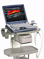
GE Logiq e Ultrasound
Good Things Come in Small Packages
Cutting-edge ultrasound innovation. Within the reach of everyone,
everywhere. A compact system that raises the bar on image quality. And
makes the power of leadership LOGIQ technology available to all clinicians.
everywhere. A compact system that raises the bar on image quality. And
makes the power of leadership LOGIQ technology available to all clinicians.
Below is a generalized description of this systems technology, specifications,
features and options. The below may not reflect the features and options
available on units in our inventory.
features and options. The below may not reflect the features and options
available on units in our inventory.
The LOGIQ e is a high performance multipurpose color compact imaging system designed for cardiac, abdominal, obstetrics, gynecology, vascular, musculoskeletal, small parts, pediatric, neonatal and intraoperative applications.
Ease of Use
- From basic ultrasound scans to more advanced imaging, LOGIQ e is easy to operate.
- Manual or automatic – You have the freedom to manually optimize images, or take advantage of dedicated imaging settings by anatomy. Just scan the way you prefer.
- Create and save your own settings – Save your preferred imaging settings as you go from one patient to the next. There’s no need to re-create the settings each time.
Advanced Imaging
- LOGIQview renders images into a panoramic image up to 60cm.
- Simultaneous Split Screen shows two Images side-by-side for live scanning, even with and without Color and Power Doppler. Also use Split Screen for contralateral and historical comparisons.
- Power Doppler Imaging (PDI) sensitivity helps you detect slow blood flow, even in
small vessels. - Easy BD gives you the power to scan and reconstruct a volumetric image.
- B-Color lets you add color tints to grayscale to better amplify contrasts
Image Processing
- Digital Beam former
- 64 Digital Processing Channel Technology
- Displayed Imaging Depth: Minimum Depth of Field: 2 cm (Zoom and probe dependent); Maximum Depth of Field: 30 cm (probe dependent)
- Transmission Focus
- 1 – 8 Focus Points Selectable (probe and application dependent
- Focal Zone Position
- Continuous Dynamic Receive
- Focus/Aperture
- Multi-Frequency/Wideband Technology
- 256 Shades of Gray (VGA)
- Adjustable Field of View (FOV)
- Image Reverse: Right/Left
- Image Rotation: 4 steps Rotation: 0°, 90°, 180°, 270°
Key Technologies Include
- Needle Recognition
- TruScan Architecture
- TruAccess
- SmartScan
- ComfortScan
Software Options
- Easy 3D
- DICOM 3.0 Connectivity
- LOGIQ View
Hardware Options
- Battery Pack
- 3 pedal Foot Switch (IPX8)
- Docking Cart
- Simple Cart
- CWD
- USB ECG (AHA/ IEC)
Physical System Specifications
- Weight: 10.1 lbs (4.6kg)
- Console Only Dimensions: 2.49″ H x 13.88″ W x 11.71″ D (61mm H x 340mm W x 287mm D)
- Console with Handle Dimensions: 3.12″ H x 13.88″ W x 13.35″ D (76.5mm H x 340mm W x 327mm D)
- Architecture: All-digital signal processing and multibeam formation technology
- Dynamic range >199 dB
- 2-D mode line density up to 512 lines
- Up to 22,560 processing channels
- DIMAQ Integrated workstation
User Interface
Operator Keyboard:
- Alphanumeric Keyboard
- Ergonomic Hard Key Operations
- Integrated Recording Keys for Remote Control of
- Peripheral Devices and DICOM Devices
- 6 TGC Pods, with Re-mapping functionality at any depth
- Backlight keys
Console Design:
- Laptop Style
- Integrated HDD (4068)
- Wireless LAN Support
- USB ECG (AHA/ IEC) (Optional) Support
- CWD (Optional) Support
- 1 probe port with micro—connector
- Rear handle
Display Screen:
- 15-inch, high resolution color LCD
- Resolution: 1024 x 768 pixels
- Total screen area: 1280 x 1024 pixels
- Interactive Dynamic Software Menu
- Integrated Speakers
- Audio Volume Adjustment
- Open Angle Adjustable – 0 to 1600
Operating Mode
- B-Mode
- Color Flow Mode
- PDI-Mode
- M-Mode
- PW/CW-Mode
- LOGIQ view
- Available on the following probes: 12L & 8L
- Virtual Convex
- Available on the following probes: 12L & 8L
Standards Features
- High Resolution 15 inch Color LCD
- 325 Frames (15 sec) Standard CINE Memory (64MB)
- 4063 Hard Drive
- External DVD RNV storage
- Loops storage-from ‘on the fly‘ scanning and from memory
- Automatic Optimization
- Auto Tissue Optimization: ATO
- Auto CFM Optimization: ACO
- Auto Spectrum Optimization: ASO
- ACE (Adaptive Color Enhancement)
- TruAccess, Raw Data Processing
- Patient Information Database
- Image Archive on Hard Drive *
- Full M&A Calculation Package with Real Time Auto
- Doppler Calculations
- Vascular Calcs
- Cardiac Calcs
- OB Calcs and Tables
- Fetal Trending
- Multi Gestational Calcs
- Hip Dysplasia Calcs
- Gynecological Calcs
- Urological Calcs
- Renal Calcs
Hard Drive
- Internal 160 GB hard drive
- Allows storage of patient studies that include images, reports and measurements
- Storage capacity of 150,00 images with compression: color or black/white
Scanning Method & Transducers
- Electronic Convex
- Electronic Linear with slant scanning
- Convex Array
- Microconvex Array
- Linear Array
- Phase Array
Media & Peripherals
- External USB DVD-RW (standard)
- USB thermal B&W printer, Sony UPD-897 (option)
- USB thermal color printer, Sony UPD-23 MD (option)
- Bluetooth wireless printers, using HP450 printers, where available
- Wireless LAN using Linksys WUSBS4G supporting the 802.11a/blg formats, where available”
- Memory Stick
System Input/ Output
- SVGA
- USB (Footswitch, DVD-RW, video printer)
- DC Power input
Power Supply
- Voltage: 100- 240 V AC
- Frequency: 50/60Hz
- Power: Max. 130 VA with Peripherals
Applications Specific Calculations
- Abdomen
- Cardiac
- Gynecology
- Small Parts
- Obstetrics
Measurements & Calculations
B-Mode:
- Distance
- Circumference/Area (Ellipse, Trace)
- % Stenosis
- Volume
- Angle
- A/B Ratio
M-Mode:
- Tissue Depth (Distance)
- Time Interval
- Depth Difference with Time Interval & Slope
- % Stenosis
- A/B Ratio
- Heart Rate
Doppler Mode:
- Velocity
- TAMAX, TAMIN, and TAMEAN (Manual/Auto Trace)
- Two Velocities with Slope and Time Interval
- Time Interval
- Pl (Pulsatility Index)
- RI (Resistive Index)
- S/D Ratio
- D/S Ratio
- AIB Ratio
- Max PG (Pressure Gradient)
- Mean PG (Pressure Gradient)
- SV (Stroke Volume)
- FV (Flow Volume)
- CO(cardiac output)
- Heart Rate
Structured Reporting
- DICOM-Cardiac Structured Reporting
- DICOM Vascular Structured Reporting
- DICOM-OB/GYN Structured Reporting
Transducers Ports
- One active port connectors
- Frequency range: 2.0 —10.0 MHz
Transducers/ Probes
- Linear Array 4-13 MHz*
- Intraoperative I Hockey Stick 4-10 MHz
- Curved Array 2—5 MHz
- Micro-Convex 4-10 MHz
- Phased l Sector Array 1.5-4 MHz
- Endocavity 4-10 MHz
- Veterinary (Equine) 4-10 MHz
Cardiac Transducer/ Probe
Adult Cardiac 1.5-4 MHz*


