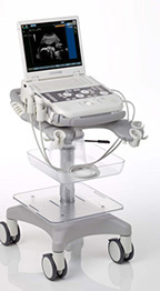
Transducers/ Probes:
- Linear Array 2.5-13 MHz*
- lntraoperative / Hockey Stick 5-13 MHz
- Curved Array 2-6 MHz*
- Micro-Convex 3.1-8.8 MHz*
- Phased / Sector Array 1-9 MHz*
- Endocavity 2.9-9.8 MHz*
- Endovaginal 2.9-9.7 MHz*
- Bi-plane Endorectal 2.9-9.0 MHz*
- Veterinary (Equine) 1-8.8 MHz
Cardiac Transducers/ Probes:
- Adult Cardiac 1.3-4.4 MHz
- Catheter 2-10 MHz*
- Pediatric Cardiac 1.4-9.2 MHz*
- Pencil 2 MHz & 5 MHz
- TEE 2.7-9 MHz
3D/ Volume Transducers/ Probes:
- Abdominal 1.9-6.4 MHz
- Endocavity 2.9-9 MHz
*Displayed MHz range includes multiple transducers
Transducers:
- VF13-5sp
- VF13-5
- VF10-5
- VF8-3
- C6-2
- C8-5
- CH5-2
- C7F2
- EV9-4
- EV9F4
- EV9-4
- EC9-4
- P9-4
- P8-4
- P5-1
- P4-2
- BP9-4
- AcuNav10F
- AcuNav8F
- V5Ms
- CW5
- CW2
Advanced technologies provide superior image quality, application versatility and confident diagnosis.
Key Technologies Include:
- Dynamic TCE tissue contrast enhancement technology
- Clarify vascular enhancement (VE) technology
- SieClear multi-view spatial compounding
- TGO Tissue Grayscale Optimization technology
- QuikStart rapid boot
- fourSight 4D transducer technology
- Advanced fourSight technology
- syngo® Auto OB measurements
- Advanced cardiology quantification applications
- One button LVO contrast
- TEE and 3D TEE using syngo fourSight TEE View
- ACUSON AcuNav catheter for lntracardiac Echocardiography
- syngo Arterial Health Package*
- CartoSound Communications for use with the ACUSON AcuNav catheter
- Cadence contrast agent imaging technology**
- Live bi-plane prostate imaging
- Customizable physician summary report package
- Specialized reporting packages for Endocrinology, Follicle Assessment, OB/GYN, Vascular, Cardiology, Emergency Medicine and more
- Live dual Bi-Plane
- P4-2 Litho transducer & Lithoskop interface SW
- Battery operation
- Barcode reader
System Features:
- Fully-adjustable, 17-inch high resolution, flat panel display reduces eye strain
- Integrated handle on monitor for improved ergonomics
- Height-adjustable control panel increases user comfort across multiple users
- Lightweight, compact system architecture facilitates mobility in any care environment
- Small, lightweight transducers for optimal user comfort
- Hanafy lens technology and SynAps synthetic aperture technology provide exceptional penetration and image uniformity at depth, reducing the amount of pressure needed to examine technically difficult-to-image patients
- Single crystal transducer technology provides broad bandwidth, increased image quality and a single probe solution for adult cardiology
Streamlined Workflow:
- Knowledge-based workflow increases consistency and decreases interoperator variability
- User-customizable control panel is fast to leam and easy to use
- Flexible archiving options to CD/DVD, USB or to DICOM-enabled networks
- Barcode reader, patient search function or DICOM worklist for easy and accurate data entry
- User-definable physician summary utility supports on-system report generation including customizable letterhead, patient data, images, comments, recommendations and signature line
- Battery
Optional Technologies Include:
- Integrated Gel Warmer
- QuikStart
- Control Panel Cover
- External Battery
- Dynamic TCE Technology
- Tissue Gray Scale Optimization (TGO)
- SieClear Multi-View Spatial Compounding
- Clarify Vascular Enhancement (VE) Technology
- SieScape Panoramic Imaging
- 3D/4D
- Advanced Cardiovascular
- Spectral and Color Doppler Tissue Imaging (DTl)
- Advnced Physio Module
- CartoSound Communication
- syngo Auto OB Measurements
- Stress Echo Imaging
- syngo fourSight TEE View
- syngo Mitral Valve Assessment (MVA)
- syngo Arterial Health Package (AHP)
- syngo Velocity Vector Imaging (VVI) Technology
- syngo Auto Left Heart (Auto LH)
- Axius Edge Assisted Ejection Fraction
- lntracardiac Echocardiography (ICE) Imaging
- Live Bi-Plane Endorectal Transducer
- Cadence Contrast Agent Imaging*
- Barcode Reader
- Veterinary Imaging
- LITHOSKOP Localization
- Modularis SW Interface
- DICOM 3.0 Connectivity
- DICOM Modality Worklist
- DICOM MPPS – Modality Performed Procedure Step
- DICOM OB Structured Reporting
- DICOM Vascular Structured Reporting
- DICOM Cardiac Structured Reporting
*This feature should be utilized according to the ASE Consensus Statement “Use of Carotid Ultrasound to Identify Subclinical Vascular Disease and Evaluate Cardiovascular Disease Risk: A Consensus Statement from the American Society of Echocardiography Carotid lntima-Media Thickness Task Force, Endorsed by the Society for Vascular Medicine.”
**At the time of publication, the US. Food and Dug Administration has cleared ultrasound contrast agents only for use in LVO. Check current regulations for the country in which you are using this system for contrast agent clearance.