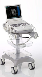
Siemens Acuson P300 Ultrasound
The Complete, Portable Patient Care Solution.
The Acuson P300 system includes advanced image optimization tools such as panoramic imaging, speckle reduction and spatial compounding, Which optimize the imaging data automatically, thus improving diagnostic confidence and supporting efficient clinical workflow.
Below is a generalized description of this systems technology, specifications, features, and options. The below may not reflect the features and options available on units in our inventory.
The ACUSON P300™ ultrasound system is a high performance compact diagnostic ultrasound that provides many advanced technologies standard with purchase, and a broad transducer suite to support individual and diverse practices; from traditional applications to specialty markets. The system is ergonomically designed for comfort and is backed by the Siemens service team for peace of mind.
- Standard package includes high-end image optimization functionalities to support traditional and specialty applications needs
- Optimized for applications in musculoskeletal (MSK), breast, small parts, cardiology, general imaging and OB/GYN
- Wide selection of Linear, Convex, Phased, Laparoscopic, Intraoperative and Endocavity probes Portability with ease
- Sleek design, easy to move and maneuver with convenient cord management system
- Integrated power supply
- Two transducer ports Support you can count on
- Backed by Siemens standard process for service and support and clinical applications
- Access to the Siemens global network of technical and application expertise to ensure optimal system performance
- A standard 2-year factory warranty* and flexible service options to meet your unique needs
Extending Technologies
- High frequency imaging
- Up to 18.0 MHz bandwidth on linear transducers to provide detailed and precise imaging, especially in superficial investigations such as MSK application
- Multiple transmission frequency to scan deeper structures without changing probes TEI
- Harmonic imaging: dedicated hardware and software processing the second harmonic frequencies improving B-mode image quality, and diagnostic confidence in technically difficult patients
- Available on all imaging transducers
- Three selectable frequencies; General, Resolution, Penetration
- Available in combination with Color Doppler (C), M-mode, Power Doppler XView
- Speckle reduction: elaborates pattern of single frame at the pixel level, eliminating speckle and noise artifacts, dynamically enhancing tissue margins, improving tissue conspicuity
- Customizable presets (X Smooth, X Detail, X Enhancement) enable real-time optimization of the image process algorithm MView
- Spatial compounding: combined contributions of standard and steered ultrasound beams to optimize image quality and improve detection of anatomical structures
- Up to 15 angles of view within the same image TPView
- Trapezoid imaging: enlarged field of view on all linear probes, allowing scanning of extended structures without losing resolution
- Specially suited for breast, vascular, musculoskeletal, and thyroid applications VPan
- Panoramic view: dedicated software merging multiple B-mode images into one panned image displayed on the screen in real-time; auto fit of composite, image zoom, merging level, frame marker, colorize, and distance measurement
- Extended field of view supports visualization of the entire organ. Areas of interest such as musculoskeletal and lesions associated with abdominal, breast and small parts can be more easily studied
The ACUSON P300 ultrasound system is specifically designed for the following applications:
- Abdominal (Adult, Pediatric, Neonatal)
- Adult Transcranial
- Breast
- Cardiac (Adult, Pediatric, Neonatal)
- Emergency
- General Imaging
- Gynecology
- Intraoperative/Interventional
- Musculoskeletal
- OB/GYN
- Small Parts
- Vascular
Physical System Specifications
- Weight: 9 kg [approx 19 lbs (without OEM’s)]
- Closed Dimensions: 35 (L) x 18 (H) x 49 (D) cm
- Working Dimensions: 35 (L) x 43 (H) x 49 (D) cm
- 15″ XVGA LCD monitor (1024×768 aspect ratio)
User Interface
- Ergonomically designed and illuminated for ease of use
- Full alphanumeric QWERTY keyboard
- Logical grouped controls
- Customizable keys
- Dedicated technology buttons; 20, M-mode, C, PW, CW
- Dedicated function keys; Start/End exam, Menu, Probe/User preset selection, Archive review
Operating Modes
- 2D (B-Mode)
- B-Mode steering
- B-Mode Auto Adjust
- Colorize 2D, M-mode and PW, CW
- PW, CW Doppler
- Non-Imaging CW
- -PW/CW Doppler Auto Adjust
- C (CoIor Doppler)
- TEI (Harmonic Imaging)
- TPView (Trapezoid Imaging)
- VPan (Panoramic Imaging)
- Bidirectional Power Doppler
Power Supply
- Voltage operative range: 100 – 240 V
- Working frequency range: 50 – 60 Hz
- Power consumption: < 250 VA
Battery
- Two removable Li-ion batteries
- Operating time: 1 hour and 20 min
- Recharging time: 100% = 3 hours and 30 min
- After 300 cycles remains 80% of maximum charge
- Nominal operating voltage is 14.4V
- Capacity: 6.6A
- Power: 95W
Cart Design
- Dimensions: 51 (W) x 84406.6 (H) x 65.4 (D)cm
- Weight: 24 kg
- Ergonomic and compact for easy maneuverability
- Four probe holders plus one Pencil probe holder
- One gel holder
- ECG cable holders
- Probe and ECG cable management system
- Multi-directional wheels with breaking mechanism and locking levers
- Height adjustable for maximum ergonomics
- On-board peripheral storage
- Foot rest for added comfort
- Rubber bumpers to prevent wall contact
Connectivity
- I/Os connectors
- Serial RS-232
- LAN RJ45
- 3 USB (for image transfer)
Dedicated Connectors
- Audio Input/output (Stereo)
- ECG input
- Double Foot Switch
- External trigger input
Image File Formats
- Standard output: BMP, JPEG, PNG, AVI
- Native and DICOM
- Clips characteristics
- AVI Codec: Microsoft® MPEG4-V2 (highly compressed), MS-WMV9 (improved compatibility) and MS-Video1 (low level compression)
- Still frames: lossy compressed (about 70% of quality)and not compressed : BMP, JPEG, PNG
- Graphic overlays
- Reports
Patient Study Management
- Internal hard drive for patient data management
- USB Data transfer
System Applications & Reporting
- Abdomen
- Emergency Medicine
- General Imaging
- Intraoperative/ Interventional
- Pediatrics
- Renal
- Veterinary Imaging
Pain Management Imaging
- Musculoskeletal (MSK)
- Anesthesia
- Orthopedic
- Podiatry
Women’s Imaging
- Breast Imaging
- Advanced OB (Option)
- Gynecology
- Obstetrics
- Reproductive Medicine (IVF)
Cardiac Imaging
- Adult Echo
- Advanced Cardiac Software (Option)
- Cardiac Screening/ Survey
- ECG
Small Parts
- Breast
- Testicle
- Thyroid
Vascular Imaging
- Arterial
- Carotid
- Transcranial
- Venous
Structured Reporting
- DICOM-Cardiac Structured Reporting
- DICOM- OB/GYN Structured Reporting
Transducer Ports
- Also supports advanced transducer technologies including fourSight 4D transducer technology
- Electronic transducer selection
- Easy-reach access to all transducer ports
- Two active port connectors
- A suite of 13 transducers that include Curved, Linear, Phased Array and 2 intraoperative probes
- Frequency Ranges: 1.0-18.0 MHz
Transducers/Probes
- Linear Array 4-18 MHz*
- Intraoperative/ Hockey Stick 4-13 MHz
- Curved Array 2-9 MHz
- Micro-Convex 3-9 MHz
- Laparoscopic 4-13 MHz*
- Phased/ Sector Array 1-11 MHz*
- Endocavity 3-9 MHz*
- Veterinary (Equine) 3-9 MHz
Cardiac Transducers/ Probes
- Adult Cardiac 1-4 MHz*
- Pencil 2 MHz & 5 MHz
- Pediatric Cardiac 4-11 MHz*
*Displayed MHz range includes multiple transducers
Transducers:
- LA522
- LA435
- LA523
- IOE323
- CA431
- CA123
- PA230
- PA023
- PA122
- EC1123
- LP323
- 2 MHZ CW
- 5 MHz CW


