Esaote MyLab Alpha Ultrasound
Top Performance In Action
MyLab Alpha, with eHD Technology focused on real diagnostic value has been designed to offer Top Performance Ultrasound and high dynamism in very reduced size and weight.
Includes 5-year standard warranty
Below is a generalized description of this systems technology, specifications, features, and options. The below may not reflect the features and options available on units in our inventory.
Imaging Processing |
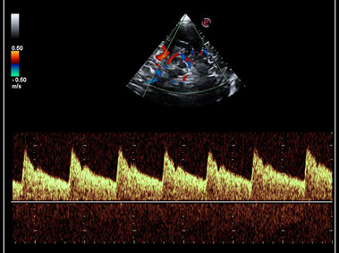
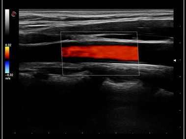
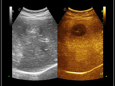
| Esaote offers many technologies for imaging enhancement. With TEI™ the harmonic signal is fully preserved without degradation of acoustic information. MView and XView improve the quality of ultrasound images by reducing the presence of artifacts, shadowing and speckle. |
|
Intelligent Software |
| eTouch™: |
|
| SmarTouch: |
|
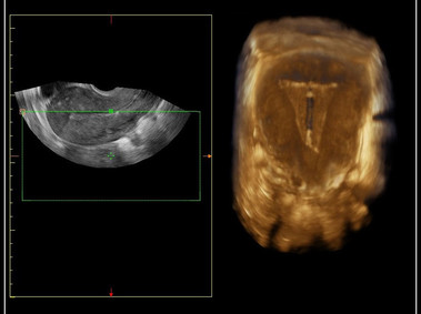
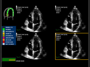
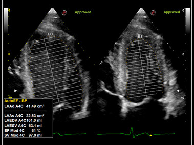
| XStrain4D: |
|
| RFQIMT – RFQAS : |
|
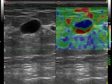
| ElaXto: |
|
Physical System Specifications |
|
Monitor
Touch Screen
|
System Overview |
|
Control Panel |
|
Enhanced Image Processing |
|
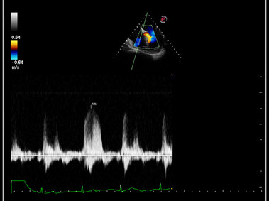
Operative Modes |
|
Archiving Capabilities |
|
Data Export |
|
DICOM Connectivity |
MyLabAlpha system supports the following DICOM service classes:
|
Standard Measurement/Reporting Packages |
|
Clinical Applications |
|
Transducer types |
|
Transducers/Probes |
|