Mindray M7 Ultrasound
Expanding the Envelope of Performance and Flexibility
The Mindray M7 epitomizes the optimal combination of advanced imaging and miniaturization technologies. Mindray research and development engineers employ the System On Chip (SOC) design within the M7 ultrasound system. SOC enables complex technologies to be built into the M7’s compact laptop style chassis. Medical devices built around SOC design are energy efficient and highly reliable.
The M7’s exceptional image quality, user experience, and versatility have expanded the performance and flexibility envelope for hand carried ultrasound imaging systems.
Below is a generalized description of this systems technology, specifications, features, and options. The below may not reflect the features and options available on units in our inventory.
Expanding the envelope of performance and flexibility
The Mindray M7 epitomizes the optimal combination of advanced imaging and miniaturization technologies. Mindray research and development engineers employ the System On Chip (SOC) design within the M7 ultrasound system. SOC enables complex technologies to be built into the M7’s compact laptop style chassis.
Medical devices built around SOC designs are energy efficient and highly reliable.
The M7’s exceptional image quality, user experience, and versatility have expanded the performance and flexibility envelope for hand carried ultrasound imaging systems.
- Compact, lightweight design enables convenient access to all point of care environments
- 15″ high resolution TFT LCD display with 170° viewing angle
- Fast power up
- Superior computing power: instantaneous response to user command and short cycle time
- Sealed surface for easy infection control
- iTouch : one button image optimization
- User programmable exam presets: quick start and consistency
- iStation : on board workstation for patient information management and connectivity
- System On Chip (SOC): enables high end system technologies in an extremely compact design
- Octal-beam imaging technology: provides excellent temporal resolution
- iClear : speckle suppression technology
- iBeam : spatial compounding
- iZoom : enables accurate viewing of image for user from distance
- iScape : panoramic imaging
- iNeedle : needle visualization technology
- TDI: tissue Doppler imaging
- IMT: auto-measurement of carotid intimae-thickness
- Full-featured DICOM function: network, worklist, MPPS, query/retrieve, structure reports
- Excellent system performance across a wide range of applications
- User selectable setup, from dedicated specialty to full service
- System ergonomic and utility: hand-carried to full service cart with external display
- Wired and iRoam, wireless, data transfer and connectivity
- Optional system cart: height adjustable, supports expanded functionalities, peripherals and storage
- System-On-Chip (SOC): enables high end system technologies in an extremely compact design
- Octal-beam imaging technology: provides excellent temporal resolution
- Innovative implementation of multiple tissue harmonic imaging technologies: improves image quality
- iZoom: enables accurate viewing of image for user from distance
- Cine compare: innovative workflow tool for measurement and report
- IMT: auto-measurement of carotid intima thickness
- TDI: tissue Doppler imaging
- FreeXros: anatomical M mode
- 4D: real-time 3D imaging
- iClear: speckle suppression technology
- iBeam: spatial compounding
- iScape: panoramic imaging
- iNeedle: needle visualization technology
- iTouch: single button image optimization
- iStation: on board workstation for patient information management and connectivity
Imaging Modes:
- 2D
- 3D/4D
Applications:
- Abdominal
- Anesthesia
- Breast
- Cardiac
- Emergency Medicine
- General Imaging
- Intraoperative
- MSK
- OB/GYN
- Pediatrics
- Neonatal
- TCD
- Small Parts
- Superficial
- Vascular
- Venous
- TEE
Features:
- M-Mode
- Color M-Mode
- PW Doppler
- CW Doppler
- Color Doppler
- Power Doppler
- DICOM
- Auto Optimization
- Speckle Reduction
- Compound Imaging
- Tissue Harmonics
- HPRF,
- Panoramic
- Needle Visualization
- IMT
- Stress Echo
- Anatomical M-Mode
- Tissue Doppler
Video & Output Options:
- S-Video
- Ethernet
- Audio
- USB
- Composite Video
- DVI
Monitor Resolution:
- 1024×768
Dimensions:
- 14 lbs, 14″ x 14″ x 3″
Export Options:
- DICOM
- JPG
- BMP
- RAW
- USB
USB Ports:
- 2
DICOM Options:
- Store
- Worklist
- MPPS
- Structured Reports
- Query/Retrieve
PC Export Formats:
- AVI
- JPG
- BMP
Wireless:
- Yes
Power (USA):
- 100-240V, 50/60 Hz
HDD Size:
- 160GB
Max Cine Memory:
- 8830 Frames
Maximum Depth (cm):
- 30
Max Frame Rate (FPS):
- 643
Accessories:
- Printer
- Footswitch
- DVD-Recorder
- Cart
- Second Battery
- Wireless Adapter
- VCR
- CD/DVD R/W
Specialty applications:
- Focused Assessment with Sonography in Trauma (FAST)
- Gallbladder
- Abdominal Aortic Aneurysm (AAA)
- Cardiology
- Focused cardiac
- Foreign bodies
- OB/GYN
- DVT (Deep Venous Thrombosis)
- Renal evaluations
- Vascular access
- Renal evaluations
- Vascular access
- Line placement
Anesthesia:
- Ultrasound-assisted procedural guidance applications
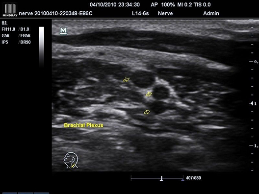
- Peripheral Nerve Blocks
- lnterscalene
- Supraclavicular
- lnfraclavicular
- Musculocutaneous
- llioinguinal/lliohyposgastric
- Femoral
- Sciatic
- Popliteal
- Saphenous
- Epidural/Spinal
- Vascular Access
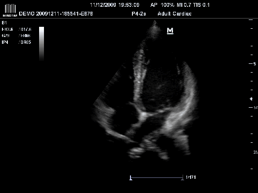
Cardiac
- Stress Echocardiograohy:
- Fast, intuitive, user friendly work flow
- 13 factory default stress protocols covering: Exercise, Dobutamine, and Ergometer
- Unlimited user defined protocols with up to 12 stages per study
- Pause function
- Easy global clip edit function
- Wall motion scoring protocols ASE 16, ASE 17
Transesophgeal Echo:
- Motor driven l Forward/reverse and lateral flexion l 180 degree scan plane I 180 degree scan plane indicator
Vascular Access
- Easily connects to your work station, giving you the ability to access essential patient reporting directly on the ultrasound system
- Access to direct applications as diverse as inputting data and editing the patient report from the workstation through the M7 while never leaving the patient’s side
- Provides immediate connectivity, bringing the user in touch with patient data streams such as electronic patient reports, at a single location, without compromising the workflow
Vascular Imaging Applications:
- Carotid exam
- Upper and lower extremity arterial exam
- Venous duplex
- Vascular Access
- Line placement
- TDC
- ABD vascular
- Comprehensive reporting
Curved Transducers/ Probes:
- C5-2s
- 6C2s (Micro)
- 4CD4s (4D)
Linear Transducers/ Probes:
- L14-6s
- L14-6ns
- 7L4s
- L7-3s
- L12-4s
- 7LT4s
Phased Transducers/ Probes:
- P4-2s
- P7-3s
- P12-4s
Endocavity Transducers/ Probes:
- V10-4bs
- V10-4s
CW (Pencil) Transducers/ Probes:
- CW2s
- CW5s
Transesophegeal Transducer/ Probe:
- P7-3ts